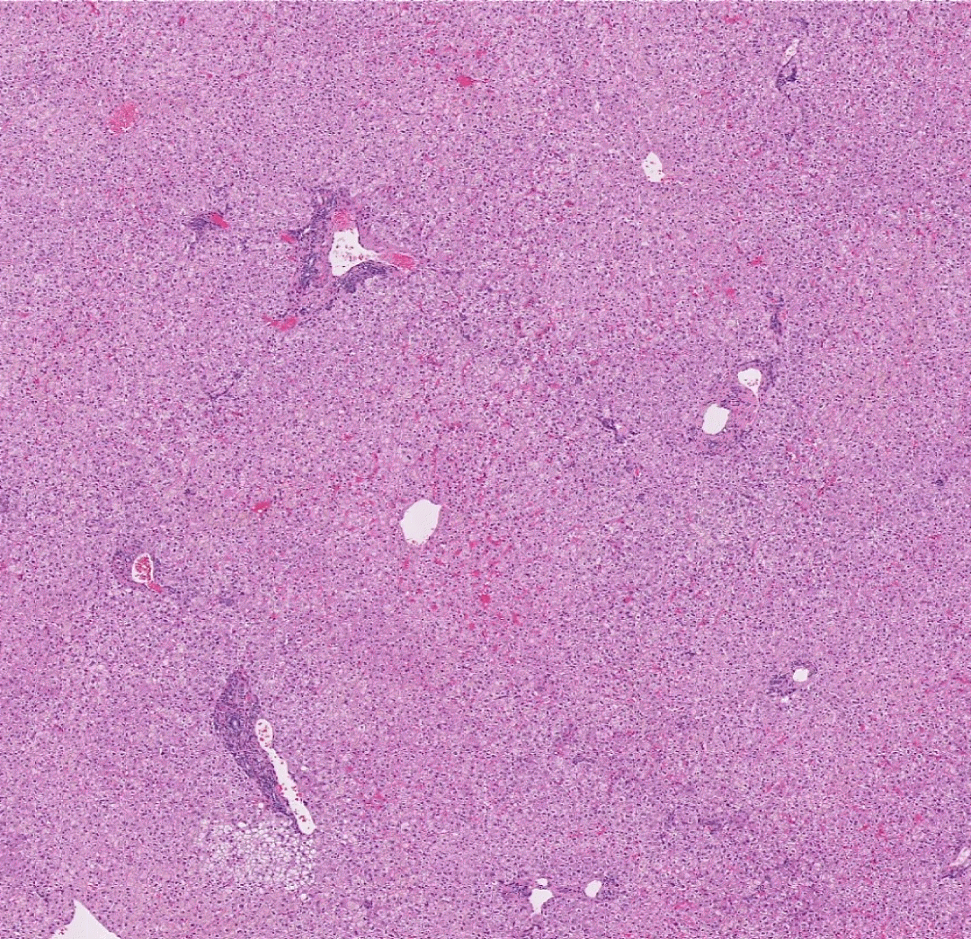


Refresh your memory of the architecture of the liver lobules. The lobules are hexagonal with portal tracts (or "triads") at the periphery and a "central" vein.
Use the buttons above to show/hide an overlay of this architecture. Move your mouse over the portal tracts and central veins to magnify them.
What 3 structures make up a portal triad?
How does the blood flow in the lobule (e.g. from where to where)?
In a hypovolemic patient, which area of the lobule would be most affected and why?
When you are ready. Press the "Return" button to go back to the folio.
The portal tracts (triads) are made up of a bile duct, a vein, and an artery. Because of the plane of section, not every corner may have an identifiable portal tract. Blood from the portal venous and hepatic arterial blood supply enters from the portal triad into the penetrating vessels at the periphery of the lobule. It mixes and then travels from the periphery, through the sinusoids where it perfuses the hepatocytes, and exits into the central vein. This path makes the area around the central vein most prone to ischemic injury.
![]()