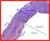
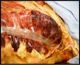
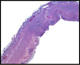
This infart is located in the watershed area between the superior and inferior mesenteric arteries and like the infarcts in the brain, point to a low flow state, as you might find in heart failure. The histology shows hemorrhage, necrosis of the wall, and focal neutrophilic infiltrate.

Here is an image of the large intestine (near the splenic flexure) of a patient who went through a period of prolonged hypoperfusion prior to death.
Can you distinguish between the normal and diseased colon on the histologic sections.
This is a similar to the process you saw in the patient with hypoperfusion injury to the brain.
 Click on the lightbulb to show the blood flow to the splenic flexure.


