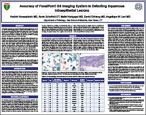
Accuracy of FocalPoint GS Imaging System in Detecting Squamous Intraepithelial Lesions
Hedieh Honarpisheh, MD, Kevin Schofield, CT, Malini Harigopal, MD, David Chhieng, MD, Angelique W. Levi, MD
Department of Pathology, Yale School of Medicine, New Haven, CT, USA
ABSTRACT
Introduction: Recently, the Food and Drug Administration approved the use of the location guided imaging system FocalPoint GS (FPGS) to assist in the primary screening of SurePath Papanicolaou (Pap) tests. A few studies have demonstrated a significant decrease in screening time and a substantial increase in the detection of squamous abnormalities. The objective of the current prospective study is to analyze the accuracy of FPGS in detecting squamous intraepithelial lesions in the clinical setting.
Materials and Methods: All SurePath Pap tests being evaluated with the assistance of FPGS during 2011 were included in the current study. False negative cases that were discovered during quality control (QC) review were retrieved. The majority of the cases that were selected for QC review were high-risk i.e. cases with previous cervical abnormalities and/or positive HPV co-testing. Only false negative cases with the final diagnosis of low-grade squamous intraepithelial lesion (LSIL) or above were included. The original 10 fields of view (FOV) were then reviewed by a senior cytotechnologist to determine if any abnormal cells were present in any of the original 10 FOVs.
Results: A total of 66,863 SurePath Pap tests were evaluated with the assistance of the FPGS imaging system during the study period. A total of 9,762 (14.6%) underwent full manual QC review. Among these cases, 105 (1.1%) cases were reclassified as LSIL or above. No abnormal cells were present in the original FOVs in 15 (14.3%) out of the 105 false negative cases. All 15 cases were reclassified as LSIL; 7 were subsequently confirmed histologically; 2 additional cases tested positive for high-risk HPV. No HSIL cases were missed by FPGS imaging system.
Conclusions: On prospective analysis, based on the results of QC review of 9,762 cases, only a small number (15/9,762; 0.15%) of abnormal squamous cells/cell clusters were not presented in the 10 FOVs by the FPGS imaging system. It is reasonable to conclude that the FPGS imaging system is relatively sensitive in detecting clinically relevant squamous cell abnormalities.
©2012 Yale Department of Pathology. All rights reserved.
Any redistribution or reproduction of part or all of the contents in any form is prohibited. You may not, except with express written permission of the author or the Department of Pathology, distribute or commercially exploit the content, nor may you transmit it or store it in any other website or other form of electronic retrieval system, including use for educational purposes.
