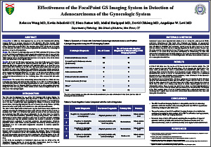
Effectiveness of the FocalPoint GS Imaging System in Detection of Adenocarcinoma of the Gynecologic System
Rebecca Wong MD, Kevin Schofield CT, Elena Ratner MD, Malini Harigopal MD, David Chhieng MD, Angelique W. Levi, MD
Department of Pathology, Yale School of Medicine, New Haven, CT, USA
ABSTRACT
Introduction: In 2008, the FDA approved the use of the BD FocalPoint GS (FPGS) Imaging System for primary screening of BD Surepath (SP) Pap smears. Data regarding the impact of imaging systems of the FPGS imaging system in the detection of adenocarcinomas is limited (2-4). The objective of the current study was to evaluate the effectiveness of FPGS in the detection of glandular abnormalities in cervico-vaginal specimens using the SP preparation.
Design: We reviewed the cytologic diagnoses all FPGS evaluated SP processed Pap tests with histologic confirmation of gynecologic adenocarcinoma over a two year time period. Cases originally interpreted as NILM were re-screened, and the ability of the FPGS imaging system to display atypical glandular cells in representative fields of view (FOV) was assessed in false negative cases.
Results: Of a total of 155 cases obtained over a period of 26 months, 72 of these cases (46%) were interpreted as atypical glandular cells (AGC 37 [24%]), adeno-carcinoma (23 [15%]), atypical squamous cells (ASCUS 9 [6%]), or a squamous lesion (LSIL 2 [1%]; HSIL 1 [0.6%]) on the SP slide. Eighty-two cases (53%) were interpreted as NILM and 1 case was interpreted as unsatisfactory. Four cases initially reported as NILM were found to contain atypical glandular cells on retrospective review. All of these cases contained atypical glandular cells in at least 1 of 10 fields of view (FOV) selected by the FPGS. The majority of the false-negative cases (3 of 4 cases) derived from endometrial adenocarcinoma and the remaining case from endocervical adenocar-cinoma.
Conclusions: The results of the current study demonstrated that the FPGS was able to identify and present glandular cell abnormalities in the FOV. This finding suggests that FPGS was effective in identifying atypical glandular cells in specimens containing malignant glandular cells. Reasons that may contribute to the false-negative diagnoses during the initial screening with FPGS include a low number of atypical cells on the slide and subtle cytologic atypia.
©2012 Yale Department of Pathology. All rights reserved.
Any redistribution or reproduction of part or all of the contents in any form is prohibited. You may not, except with express written permission of the author or the Department of Pathology, distribute or commercially exploit the content, nor may you transmit it or store it in any other website or other form of electronic retrieval system, including use for educational purposes.