Utility of Carbonic Anhydrase-IX in the Diagnosis of Metastatic Renal Cell Carcinoma by Fine-Needle Aspiration Biopsy
Xiaoling Guo, Berrin Ustun, Adebowale J. Adeniran, David Chhieng, Angelique W. Levi
Department of Pathology, Yale School of Medicine, New Haven, CT, USA
ABSTRACT
Background:
Expression of the commercially available antibody against Carbonic anhydrase-IX (CAIX) has been shown to be useful in the diagnosis of metastatic conventional renal cell carcinoma (cRCC) in surgical pathology specimens, but studies demonstrating its utility in cases of metastatic cRCC from cell-block sections of aspiration biopsies are limited. The objective of the current study was to compare CAIX expression with other immuno-histochemical markers routinely used in the diagnosis of metastatic cRCC (CD10, and Vimentin) in cell block material from fine-needle aspiration biopsies (FNABs), and to assess its utility in confirming the diagnosis of metastatic cRCC in cytological specimens.
Design:
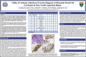
Thirteen metastatic cRCC specimens from 12 patients were immunostained with CAIX, CD10, and Vimentin. The immunoreactivity results were compared. Immunohistochemistry (IHC) staining was performed and immunoreactivity was graded as 0, no tumor cells immunoreactive (IR); 1+, 1% to 25% IR; 2+, 25% to 50% IR; and 3+, greater than 50% IR.
Results:
Eleven of 13 (85%) cases showed 3+ strong and diffuse membranous staining with CAIX; 12 of 13 (92%) cases showed similar 3+ IR with CD10; and 9 of 12 (75%) cases showed 3+ strong and diffuse cytoplasmic IR for Vimentin. In 1 case where CAIX IR was 1+, both CD10 and Vimentin showed 3+ IR; while in the other case where CAIX IR was 1+, CD10 showed 0 IR and Vimentin showed 1+ IR. Morphologically, 12 of 13 metastatic cRCCs showed small to intermediate sized nuclei with relatively inconspicuous nucleoli, and 1 case showed larger nuclei with prominent nucleoli. In the 2 cases where CAIX showed 1+ IR, the tumor cells were intermediate in size with inconspicuous nucleoli. In the 1 case with prominent nucleoli, both CAIX and CD10 showed 3+ IR, while Vimentin showed 1+ IR.
Conclusions:
The majority of cases in this study showed strong and diffuse expression of CAIX, and with the exception of 1 case the expression of CD10 and Vimentin was comparable. This study suggests that CAIX is a useful marker in addition to CD10 and Vimentin to confirm the diagnosis of metastatic cRCC in cytological cell block material from various anatomical sites.
©2012 Yale Department of Pathology. All rights reserved.
Any redistribution or reproduction of part or all of the contents in any form is prohibited. You may not, except with express written permission of the author or the Department of Pathology, distribute or commercially exploit the content, nor may you transmit it or store it in any other website or other form of electronic retrieval system, including use for educational purposes.