

Fungal
Viral
Bacterial
Normal


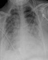
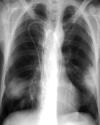
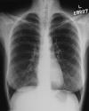
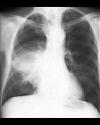
Compare the four chest radiograms. Click the icons to enlarge the image and drag them to the proper description.
Concentrate on the following questions:
1) Is (are) the lesion(s) unilateral or bilateral? It it a focal or diffuse process?
2) Does it involve one lobe or multiple lobes?
3) If they involve a single lobe, does it involve the entire lobe or only a part?
Does that help you determine the likely etiology for each x-ray?
In the case of invasive fungal pneumonia, the chest film demonstrates multiple poorly marginated areas of peripheral homogeneous opacification. Frequently these areas will become more nodular over time. The pattern is focal, but with multiple (at least 2) lesions.
In the X-ray of H1N1 pneumonia, there is bilateral consolidation (concentrated in the lower lobes) with ground glass opacity. Ground glass opacity is non-specific, but represents edema either of the interstitium (i.e. widening of the alveolar walls) or the alveoli. As with most viral pneumonia, all lobes are affected, providing a diffuse pattern of injury.
The lobar pneumonia show diffuse consolidation of the right middle lobe (hence the term "lobar" pneumonia). This pattern is diffuse, but only through one lobe.
Note that two of the patients have extensive "wiring" and/or are intubated, suggesting that they are very ill.