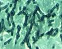


Fungal
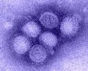
Viral
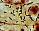
Bacterial


Here are the three causative organisms for the cases of pneumonia you have just seen.
Can you see any of these organisms in the microscopic images. If not, why not?
Is S. pneumonia encapsulated? Does that impact its pathogenicity?
Viruses are much too small to be seen by routine histology. However, many viruses induce cytopathic effects on cells that are visible (called viral inclusions). Influenza virus, however, does not. Note the hemagglutinin spikes on the surface of the virus in this electron micrograph. Influenza is categorized by the type of hemagglutinin (H) and neuraminidase (N) proteins present (hence H1N1).
Fungus does not stain will with H&E, although if you look closely in the lower left of the fungal H&E you can make out the outlines of the aspergillus. To see fungus clearly you can use either a silver stain (Grocott) or, in some cases, a Periodic acid-Schiff (PAS) stain.
Strep pneumonia is a Gram-positive diplococcus (in tissue) that can (barely) be detected using a tissue Gram stain. S pneumonia has a polysaccharide capsule which prevents phagocytosis by macrophages; non-encapsulated Streptococcus pneumonia are not pathogenic.
The incidence of S. pneumonia infections has declined with the use of the pneumococcal vaccine. Another cause of lobar pneumonia is Klebsiella.