

Fungal
Viral
Bacterial
Normal


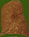
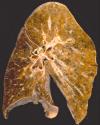
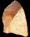
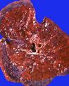
Now compare some gross specimens of the same conditions.
Also consider how these gross specimens might match up with the radiograms (although they are clearly taken from different patients than the X-rays).
Determine if the disease process is diffuse or limited to one lobe.
Note the location of the lesion in the top image. Does that tell you anything about the type of infectious process?
How might the infections process have spread in the middle specimen?
In the case of invasive fungal pneumonia, infection has invaded from one lobe into another across the lobal fissure. That is consistant with the "invasive" nature of Aspergillus (we will discuss the types of Aspergillus infections in more detail on the next page).
The lobar pneumonia shows "gray hepatization" of the lower lobe with relative sparing of the upper lobe. In lobar pneumonias, inflammatory cells fill the lobules and then spread, via the airways, to adjacent lobules, eventually filling the whole lobe.
The viral pneumonia shows diffuse consolidation and hemorrhage as would be found in cases of diffuse alveolar damage.