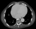
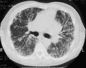
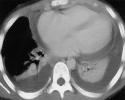
Pleural effusions (colored blue) occur in 30-50% of lupus patients bout only 5% of patients with systemic sclerosis. The etiology is not entirely clear, but may be a consequence of trapping of immune complexes in the pleural fluid filtrate. Pericardial effusions are also relatively common (colored yellow). The normal and lupus images are taken from the lower lung fields (note the presence of the heart and the lack of bronchi or great vessels). The either/both image is taken towards the middle of the lung (note the presence of the left and right bronchi which show up as dark elongated holes in the center of the image).
Usual Interstitial Pneumonia (UIP) leading to pulmonary fibrosis is a common finding in both scleroderma and lupus. Note the decreased lung volumes and prominent reticular interstitial markings particularly in the base of the lung at the periphery. Late stages show diffuse "honeycombing", characterized by multiple cystic spaces surrounded by fibrotic bands. Ground glass opacities are usually a minor component unless there is active disease.


Normal
Both
Lupus
Compare these CT scans. Where are they taken (i.e. top, middle, lower lung fields)?
Where is the pathology and how would you describe the radiologic findings?