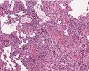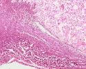


Normal
Both
Lupus
The "Lupus" image shows extensive fibrosis attached to the pleural lining of the lung. Note the lung parenchyma is relatively normal.
The "Both" image shows UIP, the type of pulmonary fibrosis that can occur in either disease. Note the interstitial fibrosis here is at the periphery of the lung and shows interstitial scarring, honeycomb changes and fibroblast foci, with only mild chronic inflammation. Early in the disease, the changes can be patchy with different areas exhibiting a spectrum of fibrosis from normal to active to completely fibrotic.


These microscopic images match the CT scans and gross images.
Can you characterize the pathology and determine the correct image order?