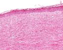


Normal
Lupus
S. Sclerosis
Compare the H&E sections of the skin. Drag the icons to the appropriate slot.
Look for both changes in the epidermis and dermis. Knowing something about systemic sclerosis, what do you think the skin will show?
Is there Inflammation? Necrosis? Apoptosis? Fibrosis?
![]() In the lupus skin, note the degeneration of the basal layer of the epidermis, edema of the dermis, inflammation along the dermal/epidermal junction, and vasculitis. Also note the apoptotic keratinocytes (highlighted in the image below). Sunlight exacerbates the erythema seen in skin of lupus patients. Some researchers (see the pdf) have speculated that UV radiation induces the apoptosis of keratinocytes, and that lupus patients have defective macrophage clearance of these apoptotic keratinocytes. This results in an increased burden of nuclear antigens that stimulated lymphocytes, ultimately leading to the production of anti-nuclear antibodies (ANAs).
In the lupus skin, note the degeneration of the basal layer of the epidermis, edema of the dermis, inflammation along the dermal/epidermal junction, and vasculitis. Also note the apoptotic keratinocytes (highlighted in the image below). Sunlight exacerbates the erythema seen in skin of lupus patients. Some researchers (see the pdf) have speculated that UV radiation induces the apoptosis of keratinocytes, and that lupus patients have defective macrophage clearance of these apoptotic keratinocytes. This results in an increased burden of nuclear antigens that stimulated lymphocytes, ultimately leading to the production of anti-nuclear antibodies (ANAs).